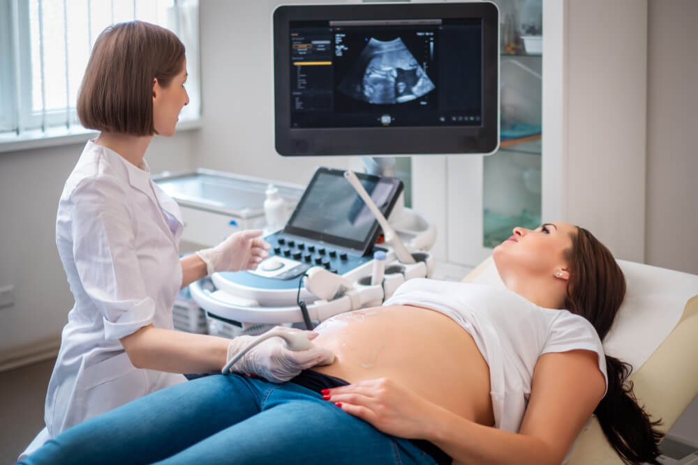
Transabdominal ultrasound
Transabdominal ultrasound in nagpur is the method through which medical imaging is produced whereby high frequency sound waves create images from inside abdominal organs and structures, and in this process, the probe or transducer is applied over the abdominal wall to the patient.
– The gel is used on the transducer to the ensure that the image produced is of good quality.
– Sound waves are transmitted through the abdominal organs or structures
-The reflected sound waves converted into images form on a screen
What is Transabdominal Ultrasound?
A non-invasive medical imaging technique that makes use of high frequency sound waves to create images of the abdominal organs and structures
What is Transabdominal Ultrasound used for?
Evaluates the liver, gallbladder, pancreas, kidneys, or a spleen
Can identify any conditions such as gallstones, liver disease, and kidney stones
It can be monitored for fetal development during pregnancy
It guides biopsies or tumor treatments
Abdominal pain or discomfort evaluation
Preparation for Transabdominal Ultrasound
No special preparation is necessary
– May require the patient to fast for 8-12 hours before the procedure
– Patient should come dressed in loose clothing avoiding tight cloth around the abdomen
Advantages
– It is non-invasive and painless.
– There is no radiation exposure.
– Real-time imaging leads to quicker and quicker diagnosis
– Easy, compared with other imaging modalities and relatively inexpensive
Limitations
– Imaging quality may be compromised in an obese patient
– Maybe not sensitive to all abdominal conditions or diseases
– Shallow penetration decreases the ability to image deep structures.
Who does Transabdominal Ultrasound
-Radiologist or a technologist in ultrasound
When to avoid Transabdominal Ultrasound
-Patients with open wound or surgical wound in the abdomen
-Patients with severe abdominal pains or discomfort
-Patients who are unable to lie on the examination table
What Is a Transabdominal ultrasound?
One of the medical imaging technologies that rely on high frequency sound waves to produce an image of abdominal organs and structures from outside the human body includes a transabdominal ultrasound. It is non-invasive or painless and thus helps in diagnosing several conditions affecting the abdomen.
How it works
This is how it works:
Placement of the transducer, also known as a probe, on the abdomen
Application of gel to enhance a quality of the images
Sends sound waves to the abdominal organs or structures
– Sound waves reflected back are a displayed as images on a screen
Transabdominal ultrasound is used to examine the following:
– Liver
– Gallbladder
– Pancreas
– Kidneys
– Spleen
– Fetus in pregnancy
It helps identify the following conditions:
– Gallstones
– Diseases of the liver
– Kidney stones
– Pancreatitis
– Abdominal masses or tumors
A transabdominal ultrasound will be considered appropriate for your case by your doctor.
Uses of Transabdominal Ultrasound:
Pregnancy Scanning:
To establish pregnancy, assess the fetus’s growth and position or verify the amniotic fluid level or the health of the placenta.
Reproductive Health:
Examine the presence of fibroids, ovarian cysts, and any abnormalities within the uterus, ovaries, and fallopian tubes.
Bladder Scanning:
Inspect the bladder for any tumor development, bladder stones, or other conditions.
Kidneys and other Pelvic Organs:
Can be applied in scouting for kidney stones or any other conditions within the urinary system.
Pelvic Pain:
Such diagnosis is done for causes of pelvic pain, which may be infections or problems with the reproductive organs.
Concerns in Post-menopausal
All such unusual symptoms or bleeding in post-menopausal women are investigated for causes.
What procedures Transabdominal ultrasound?
Procedures for Transabdominal Ultrasound.
- Abdominal ultrasound: This scans the liver, gallbladder, pancreas, kidneys, and spleen.
- Obstetric ultrasound: Determines the condition pertaining to fetal development or well-being in terms of gestation periods.
- Ultrasound of the liver: Conditions such as cirrhosis, hepatitis, and cancer of the liver.
- Ultrasound of the gallbladder: Detects gallstones and inflammation of the gallbladder.
- Kidney ultrasound: Examines kidney stones and kidney disease and cancer.
- Pancreatic ultrasound: Determined pancreatitis, pancreatic cancer, or a cysts of the pancreas.
- Spleen ultrasound: Examines enlargement or rupture of the spleen.
- Abdominal Pain Assessment: Helps evaluate the cause of the discomfort or pain felt in the abdomen.
- Mass or tumor evaluation: Evaluation of an abdominal mass and tumor.
- Follow-up Ultrasound: Conducting follow-up checks of a condition previously diagnosed by ultrasound.
At our Neurosys Multispeciality Center, we perform several key procedures including Craniotomy, which is primarily for the excision of brain tumors; V-P Shunt Surgery for treating hydrocephalus; surgeries for epilepsy; and operations targeting brain stem glioma. Beyond these, we offer a range of other neurosurgical services. If you have any questions that are not answere, please contact us through our Contact Us or Book your Appointment.
