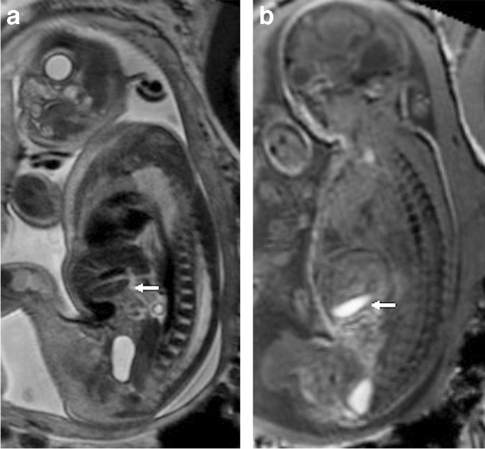
Fetal magnetic resonance imaging (MRI)
Fetal MRI in Nagpur is a non-invasive medical imaging technique that uses magnetic fields and radio waves to create precise images of the fetus at pregnancy.
Fetal MRI gives detailed resolution on the anatomy of the fetus that provides the possibility for:
1. Detection of congenital anomalies and abnormalities
2. Evaluation of fetal growth and development
3. Evaluation of placental function and umbilical cord blood flow
4. Detection of fetal distress or complications
5. Counseling about the need to perform invasive procedures like amniocentesis or fetal surgery
Especially it is a helpful in the following situation:
1. Fetal brain and spine evaluation
2. Fetal heart and cardiovascular system assessment
3. Fetal lung and diaphragm evaluation
4. Fetal gastrointestinal tract assessment
5. Detection of fetal tumors or masses
Fetal MRI is a usually conducted after 18-20 weeks of a gestation. The advantages include,
1. Radiation-free.
2. High-resolution images.
3. Non-invasive procedure.
4. Mother and fetus are not at risk.
It is however not appropriate for all pregnancies, and some of its disadvantages include the following:
1. Claustrophobia and anxiety in certain patients
2. An inability to see well in the earlier gestation periods (<18 weeks)
3. Artifacts caused by fetal motion or maternal respiration
In general, the application of the fetal MRI as a useful diagnostic tool will be helpful in assessing fetal health and detecting the development of any complications during pregnancy.
What Is a Fetal magnetic resonance imaging (MRI)?
Fetal MRI is a modality based on the principles of the strong magnetic field and effects of radio waves that produces highly defined images of the fetus in utero. The detailed high-resolution anatomy of the fetus in utero enables:
- Evaluation of fetal growth and development
- Evaluation of placental function or umbilical cord blood flow
- Detection of fetal distress or complications
- Guidance of invasive procedures such as amniocentesis or fetal surgery
It is absolutely non-invasive, non-ionizing or safe procedure for both the mother and the fetus. Normally, it is carried out after 18-20 weeks of gestation and comes into use in assessing multiple structures in the fetus such as the brain, spinal cord, heart, lungs, or gastrointestinal tract.
In particular, it is useful in the diagnosis of complex congenital anomalies, evaluation of fetal growth and development, identification of potential complications and guidance of fetal therapy and surgery.
Altogether, fetal MRI offers rich information about the health and development of a fetus, thus helping healthcare professionals in the planning of options of best care.
sleep, consciousness, and cognitive abilities.
Some common signs of neurological conditions are as mentioned below:
Pain-Associated Symptoms:
General pain, back pain, neck pain, headaches
Pain along nerve pathways, such as with sciatica and shingles
Muscle Function Abnormalities:
- Muscle weakness or tremors
- Paralysis and involuntary movements such as tics
- Walking abnormalities, clumsiness, poor coordination
- Muscle spasms, rigidity, stiffness, and spasticity
- Slow movements
Changes in Sensations:
- Numbness or tingling
- Hyperesthesia (increased sensitivity to touch)
- Loss of a sensation to touch, temperature, and pain
- Impaired proprioception sense (position of the body)
- Special Senses Alterations:
Disturbances in visions with hallucinations and partial or total loss of vision or diplopia. Also, a person may face hearing loss or tinnitus.
Key Features of Fetal MRI:
- Non-invasive: Uses magnetic fields and does not involve radiation, making it safe for both the mother and the fetus.
- Detailed Imaging: Provides high-resolution images of the fetal brain, spine, lungs, abdomen, and other organs, offering greater clarity than ultrasound in certain cases.
- Supplement to Ultrasound: Typically performed when ultrasound findings are inconclusive or if a more detailed evaluation of the fetus is needed.
Indications for Fetal MRI:
Fetal MRI is usually recommended when there is a suspected abnormality on the ultrasound, or when a more in-depth look at certain organs is required. Some common indications include:
1. Brain and Central Nervous System (CNS) Anomalies
- Hydrocephalus: Enlargement of fluid-filled spaces in the brain.
- Arnold-Chiari Malformation: A condition where parts of the brain extend into the spinal canal.
- Holoprosencephaly: A brain malformation where a forebrain fails to divide into the two hemispheres.
- Neural Tube Defects: Spina bifida or other malformations of the spinal cord.
- Cerebral Dysgenesis: Abnormal development of brain structures, such as agenesis of the corpus callosum.
2. Congenital Lung and Chest Abnormalities
- Congenital Diaphragmatic Hernia: Herniation of abdominal organs into a chest cavity due to the defect in the diaphragm.
- Cystic Lung Lesions: Pulmonary cysts or masses, such as congenital cystic adenomatoid malformation (CCAM).
3. Abdominal and Genitourinary Abnormalities
- Abdominal Wall Defects: Conditions like omphalocele and gastroschisis, where abdominal organs protrude outside the body.
- Renal (Kidney) Anomalies: Issues like polycystic kidney disease, hydronephrosis, or renal agenesis.
- Congenital Tumors: Teratomas or other fetal tumors that may be detected early.
4. Musculoskeletal Abnormalities
- Skeletal Dysplasias: Disorders affecting bone growth, such as dwarfism or bone malformations.
- Limb and Spinal Defects: Conditions like clubfoot or scoliosis that require detailed evaluation.
5. Placental and Umbilical Cord Abnormalities
- Placenta Accreta: Invasive growth of the placenta into the uterine wall.
- Umbilical Cord Abnormalities: MRI can help assess cord abnormalities that might affect fetal blood flow.
Advantages of Fetal MRI:
- Superior Soft Tissue Contrast: MRI provides a clearer view of soft tissues, making it ideal for evaluating the fetal brain and other organs.
- Non-reliant on Amniotic Fluid: Unlike ultrasound, which depends on the presence of amniotic fluid for clear images, MRI can produce high-quality images even when fluid levels are low (oligohydramnios).
- Evaluation in Complex Conditions: When conditions like fetal tumors or congenital malformations are suspected, MRI can offer more detailed information to guide treatment plans.
Safety of Fetal MRI:
- No Ionizing Radiation: Unlike X-rays or CT scans, MRI does not expose the fetus to radiation, making it a safer option.
- Magnetic Fields: MRI uses strong magnetic fields, which are generally considered safe for the fetus. However, it is usually avoided in the first trimester unless absolutely necessary due to limited data on early exposure.
Limitations:
- Fetal Movement: Since the fetus moves freely in the womb, excessive movement can make it challenging to obtain clear images.
- Limited Availability: Not all centers may offer fetal MRI, and it requires specialized expertise to interpret the results.
- Timing: While MRI is safe during the second and third trimesters, it is usually not performed in the first trimester due to limited information about potential risks to the developing fetus during that early stage.
What procedures Fetal magnetic resonance imaging (MRI)?
Procedures under fetal MRI include:
1. Fetal anatomy survey: Studies the fetal anatomy, including brain, spine, heart, lungs, and gastrointestinal tract.
2. Fetal brain MRI: It assesses the growth of a fetus’s brain in order to identify abnormalities like ventriculomegaly or cerebral malformations.
3. Fetal heart MRI: It studies the fetal cardiac structure and function to identify congenital heart defects.
4. Fetal Lung MRI: It studies the developmental process of fetal lungs and investigates anomalies like congenital diaphragmatic hernia.
5. Fetal Abdomen MRI: Assess abdominal organs of the fetus, defect like gastroschisis and omphalocele can be identified,
6. Diagnosis of Congenital Anomalies by Fetal MRI: It can be used for the diagnosis and characterization of congenital anomalies as well as management and therapy guidance.
7. Estimation of Fetal Growth and Development: It is helpful in the estimation of fetal growth to identify growth restriction or macrosomia.
8. Placental MRI: Assesses the function of the placenta and can identify abnormalities in placenta accreta or insufficiency of the placenta.
9. Fetal MRI in Maternal-Fetal Medicine: This can guide high-risk pregnancy cases and detect complications like fetal distress.
10. Research Protocols: It discusses studies on fetal development, placental function, among others, as complications in pregnancy.
These procedures require a specialized MRI machine and expertise in interpreting fetal MRI. The information they yield is very valuable to prenatal care and management.
At our Neurosys Multispeciality Center, we perform several key procedures including Craniotomy, which is primarily for the excision of brain tumors; V-P Shunt Surgery for treating hydrocephalus; surgeries for epilepsy; and operations targeting brain stem glioma. Beyond these, we offer a range of other neurosurgical services. If you have any questions that are not answere, please contact us through our Contact Us or Book your Appointment.
