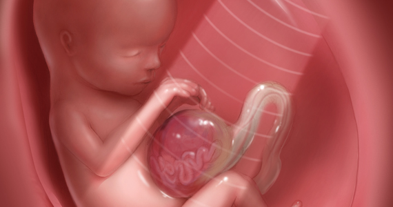
Fetal anomalies
Fetal anomalies in nagpur are those defects or abnormal development that is usually found in the fetus during pregnancy. They could be widespread and may include:
1. Heart (congenital heart defects)
2. Brain and spine (neural tube defects or hydrocephalus)
3. Limbs (club foot, polydactyly)
4. Organs (kidney, liver, lung abnormalities)
5. Gastrointestinal tract (diaphragmatic hernia, gastroschisis)
6. Genitalia (hypospadias, ambiguous genitalia)
7. Skeletal system (club foot, achondroplasia)
Many prenatal tests can detect fetal anomalies, such as:
- Ultrasound
Amniocentesis - Chorionic villus sampling or CVS
- Fetal MRI
- Fetal echocardiography
Some of these anomalies can be treated in utero, or immediately after birth, but others require lifelong management and care. It is essential for the expectant parents to be an active participant with their obstetric provider to learn how a baby’s condition may be diagnosed, what course of treatment to expect, and what the outcome will be for their baby.
What Is a Fetal anomalies?
Fetal anomalies are otherwise known as congenital anomalies. These refer to abnormal developments or birth defects that occur within the fetus during pregnancy, affecting various portions of the body such as the heart, brain, spine, limbs, organs, and gastrointestinal tract.
Fetal anomalies can be:
1. Structural: Physical abnormalities in which conditions could be heart defects, cleft lip, or another similar condition
2. Chromosomal: Genetic abnormalities, like Down syndrome
3. Metabolic: Inborn errors of metabolism, like PKU or other similar disorders.
4. Developmental: Growth and developmental defects which may include growth restriction.
Some conditions that cause fetal anomalies include:
1. Genetic mutations
2. Environmental factors including maternal infection or certain medications
3. Random errors during the development of the fetus
Some of the common fetal anomalies are as follow
1. Congenital heart defects
2. Neural tube defects (spina bifida)
3. Cleft lip and palate
4. Club foot
5. Diaphragmatic hernia
6. Gastroschisis
7. Hydrocephalus
8. Chromosomal abnormalities including Down syndrome, Trisomy 13.
Accurate early detection and diagnosis of fetal anomalies play an important role in proper management and care of the mother and the fetus during pregnancy and after birth.
Types of Fetal Anomalies
- Structural Anomalies:
- Neural Tube Defects: Conditions such as spina bifida (incomplete closure of the spinal column) or anencephaly (absence of a major portion of the brain and skull) that occur when the neural tube does not close properly.
- Congenital Heart Defects: Abnormalities in the heart’s structure, such as ventricular septal defects (holes in the heart’s walls), coarctation of the aorta (narrowing of the aorta), and transposition of the great arteries.
- Cleft Lip and Palate: A split in the upper lip and/or roof of the mouth that occurs when the tissue does not fully come together during early development.
- Abdominal Wall Defects: Conditions like gastroschisis (intestines protrude through an opening in the abdominal wall) and omphalocele (organs are outside the body but covered by a membrane).
- Limb Anomalies: Abnormalities affecting the limbs, such as clubfoot (feet turned inward) or limb reduction defects (missing or shortened limbs).
- Facial Anomalies: Abnormalities in facial structures, such as hypospadias (urethral opening on the underside of the penis) or craniosynostosis (premature fusion of skull bones).
- Chromosomal Anomalies:
- Down Syndrome (Trisomy 21): A genetic condition caused by the presence of an extra chromosome 21, leading to developmental delays and characteristic physical features.
- Edwards Syndrome (Trisomy 18): A genetic condition resulting from an extra chromosome 18, associated with severe developmental and physical problems.
- Patau Syndrome (Trisomy 13): A genetic disorder caused by an extra chromosome 13, leading to severe intellectual disabilities and physical anomalies.
- Turner Syndrome: A condition affecting females, where one of the X chromosomes is missing or partially missing, leading to various physical and developmental challenges.
- Functional Anomalies:
- Renal Anomalies: Conditions affecting the kidneys, such as agenesis (absence of a kidney), horseshoe kidney (kidneys fused together), or polycystic kidney disease (fluid-filled cysts in the kidneys).
- Gastrointestinal Anomalies: Abnormalities in the digestive system, such as esophageal atresia (esophagus does not connect to the stomach) or duodenal atresia (blockage in the first part of the small intestine).
- Neurological Anomalies: Conditions affecting the nervous system, such as holoprosencephaly (failure of the forebrain to properly divide into two hemispheres) or hydrocephalus (excess cerebrospinal fluid in the brain).
Causes of Fetal Anomalies
Fetal anomalies can result from various factors, including:
- Genetic Factors: Abnormalities in the chromosomes or genes can lead to congenital conditions.
- Environmental Factors: Exposure to teratogens (substances that can cause birth defects) during pregnancy, such as certain medications, alcohol, tobacco, or infections (e.g., rubella, cytomegalovirus).
- Maternal Health Conditions: Conditions like diabetes, obesity, or autoimmune disorders may increase the risk of fetal anomalies.
- Nutritional Deficiencies: Lack of essential nutrients, particularly folic acid, during pregnancy can lead to neural tube defects.
Diagnosis of Fetal Anomalies
- Prenatal Screening Tests:
- First Trimester Screen: A combination of blood tests and ultrasounds to assess the risk of chromosomal abnormalities.
- Second Trimester Screen: Blood tests that measure specific markers to evaluate the risk of certain conditions.
- Non-Invasive Prenatal Testing (NIPT): A blood test that analyzes fetal DNA in the mother’s blood to screen for chromosomal abnormalities.
- Diagnostic Tests:
- Ultrasound: Detailed ultrasounds can identify structural anomalies in the fetus, particularly during the second trimester.
- Amniocentesis: A procedure that tests amniotic fluid for genetic conditions, usually performed if a high risk for abnormalities is identified.
- Chorionic Villus Sampling (CVS): A test that collects tissue from the placenta for genetic analysis.
- Fetal MRI: In some cases, an MRI may be used to provide more detailed images of the fetus’s anatomy, particularly for complex anomalies.
Management and Treatment of Fetal Anomalies
Management and treatment options for fetal anomalies depend on the type and severity of the anomaly:
- Monitoring: Some mild anomalies may require close monitoring during pregnancy and after birth.
- Interventions: In certain cases, in-utero interventions may be performed to treat or manage anomalies (e.g., fetal surgery for spina bifida).
- Specialized Care: After birth, newborns with congenital anomalies often require specialized medical care, including surgery, therapy, or ongoing medical management.
- Genetic Counseling: Families may benefit from genetic counseling to understand the implications of the diagnosis, potential outcomes, and future reproductive options.
What procedures Fetal anomalies?
Fetal anomalies may thus require a variety of procedures for diagnosis, treatment, and postnatal management. Some of the common procedures include:
1. Ultrasound: Detailed ultrasound scans with the aim of visualizing the fetus and then actually identifying anomalies.
2. Amniocentesis: Sampling of amniotic fluid to a check for chromosomal abnormalities and genetic disorders.
3. Chorionic villus sampling (CVS): Cell sampling from the placenta is conducted to ascertain if the cells have a chromosomal abnormality.
4. Fetal MRI: Magnetic Resonance Imaging to the provide detailed images of the fetus.
5. Fetal echocardiography: Is a test that uses sound waves (ultrasound) to evaluate the baby’s heart for problems before birth.
6. Fetal blood sampling: Is a procedure used to diagnose, treat and monitor various fetal problems
7. Intrauterine treatment: Procedures performed directly on the fetus, such as a blood transfusions and shunt placement.
8. Fetal surgery: The surgical interventions done on the fetus, including repair of congenital anomalies.
9. Postnatal surgery: Those interventions undertaken after the birth for correction of congenital anomalies.
10. Genetic counseling: Counseling of families for a understanding the disorders caused genetically or risk assessments.
11. Prenatal consultation: Conferencing with specialists regarding diagnosis, options of treatment, and management planning.
12. Fetal intervention: Procedures aimed at treating or managing fetal anomalies, including shunt placement or radiofrequency ablation.
These procedures aid the diagnosis and management of various types of congenital anomalies, including:
Congenital heart defects
Neural tube defects
Cleft lip and palate
Club foot
Diaphragmatic hernia
Gastroschisis
Hydrocephalus
Chromosomal abnormalities
For example, a health care provider can observe and apply which procedure and with what frequency may be appropriate for a specific case.
At our Neurosys Multispeciality Center, we perform several key procedures including Craniotomy, which is primarily for the excision of brain tumors; V-P Shunt Surgery for treating hydrocephalus; surgeries for epilepsy; and operations targeting brain stem glioma. Beyond these, we offer a range of other neurosurgical services. If you have any questions that are not answere, please contact us through our Contact Us or Book your Appointment.
