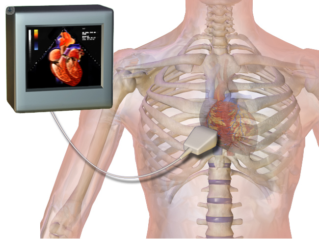
Echocardiography
Echocardiography in nagpur, also known as cardiac ultrasound or echo, is a non-invasive medical imaging test that uses high-frequency sound waves to produce detailed images of the heart and its blood vessels. It helps diagnose or monitor various heart conditions, including:
1. Heart valve problems
2. Cardiac chamber sizes and function
3. Heart failure
4. Coronary artery disease
5. Cardiomyopathy
6. Pericardial disease
7. Congenital heart defects
There are four different types of echocardiography.
1. The most common type is the transthoracic echocardiogram, which is performed with the aid of a probe on the chest to reveal an image of the heart.
2. Transesophageal echocardiogram: This uses a probe passed through the mouth or the nose to create images from inside the esophagus.
3. Stress echocardiogram: This combines ultrasound for it to assess the functioning of the heart under physical stress resultant from either exercise or medical usage of medications.
4. Contrast echocardiogram: This sort of examination would require the use of a contrast agent for quality images.
5. 3D echocardiogram: It creates a detailed 3D view of the heart.
6. Fetal echocardiogram: It is utilized for evaluation of the heart of a fetus during the womb.
Echocardiography is useful in:
- Diagnosis of heart diseases
- Monitoring Heart Functioning
- Cardiac procedure
- For the follow up of effectiveness of the treatments
- Detection of possible heart disease in those patients who have the risk factors.
This is a non-invasive, painless, and radiation-free imaging tool that provides relevant information to diagnose and manage heart diseases.
What Is a Echocardiography?
Echocardiography, often referred to as cardiac ultrasound or simply echo, is a type of non-invasive medical imaging scan. It uses high-frequency sound waves called ultrasounds to create detailed images of the heart and its blood vessels. Heart conditions may be diagnosed and monitored, including:
– Heart valve diseases
– Sizing of heart chambers and function
– Heart failure
– Ischemic coronary disease
– Cardiomyopathy
– Pericardial disease
– Congenital heart abnormalities
– Cardiac neoplasms
The procedure is performed by a cardiologist or sonographer by placing a probe (transducer) on the chest, also called a transthoracic echocardiogram; sometimes done directly through the esophagus known as a transesophageal echocardiogram. It is:
– Nonpainful
– Noninvasive
– Radiation-free
– Fast, typically 15-30 minutes in length
Echocardiography provides very valuable information that helps diagnose and monitor the management of heart conditions and can be used to assess heart function and identify other issues in the heart, direct cardiac procedures and monitor response to treatment.
Types of Echocardiography:
Transthoracic Echocardiography (TTE):
- The most common type, where the transducer is placed on the chest to produce images of the heart.
- Non-invasive, painless, and quick.
Transesophageal Echocardiography (TEE):
- Provides more detailed images of the heart, especially for detecting issues with the valves or chambers.
- A thin, flexible tube with a transducer is inserted down the esophagus (behind the heart), offering closer views.
- Requires mild sedation.
Stress Echocardiography:
- Performed while the heart is under stress, either through physical exercise (like a treadmill or stationary bike) or with medication that stimulates the heart.
- Helps assess how well the heart functions during physical activity and can detect coronary artery disease (blockages or narrowed arteries).
Doppler Echocardiography:
- A specialized type of echo that evaluates blood flow through the heart and blood vessels.
- Measures the speed and direction of blood flow to detect problems like valve dysfunctions or abnormal blood flow (e.g., regurgitation or stenosis).
3D Echocardiography:
- Provides three-dimensional images of the heart, giving a more accurate and detailed view of structures like heart valves.
- Useful in planning surgeries, particularly for valve repair or replacement.
Fetal Echocardiography:
- Used to assess the heart of a developing fetus during pregnancy, particularly if there is a family history of heart defects or other risk factors.
What procedures of Echocardiography?
Procedures which are included under Echocardiography are:
1. Transthoracic Echocardiogram, TTE: This is the most common form of an echocardiogram; a probe is placed on the chest, and it creates an image of your heart.
2. Transesophageal Echocardiogram, TEE: This is done when there is a use of a probe placed in the mouth or even through the nose to create pictures of your heart through your esophagus.
3. Stress Echocardiogram: This is used when ultrasound is used with physical stress (exercise or medication) to test for a heartbeat working capability under stress.
4. Contrast Echocardiogram: This suggests that a contrast agent is used in creating good quality images.
5. 3D Echocardiogram: It forms very detailed 3D images of the heart.
6. Fetal Echocardiogram: The procedure aids in evaluating the heart of a fetus while it is still in the womb.
7. Intracardiac Echocardiogram (ICE): A specialized probe is inserted into a vein to capture images of the heart from inside the heart chambers.
8. Intravascular Ultrasound (IVUS): It uses a small probe inserted through a blood vessel that gains images of the inner linings of blood vessels.
9. Echo Guided Intervention: Utilizing echocardiography as a guide for procedures like heart valve repair or heart valve replacement.
10. Cardiac Resynchronization Therapy (CRT) Optimization: In this process, echocardiography is utilized to optimize CRT settings on a CRT device.
At our Neurosys Multispeciality Center, we perform several key procedures including Craniotomy, which is primarily for the excision of brain tumors; V-P Shunt Surgery for treating hydrocephalus; surgeries for epilepsy; and operations targeting brain stem glioma. Beyond these, we offer a range of other neurosurgical services. If you have any questions that are not answere, please contact us through our Contact Us or Book your Appointment.
