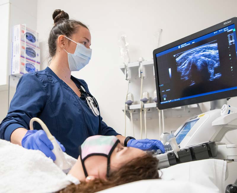
Ultrasound imaging
Ultrasound imaging in Nagpur or sonography is a diagnostic test that helps in obtaining photographs of the internal structures of the body without surgery or incision.
Main Points about Ultrasound Imaging:
– It uses sound waves for producing pictures
– It is painless, non-invasive;
– No radiation exposure
What is Ultrasound Imaging?
Ultrasound imaging – uses high frequency sound waves to produce images of internal structures; also referred to as sonography.
How does Ultrasound Imaging work?
A probe called a transducer sends sound waves into the body and they bounce off internal structures and return to the transducer that it converts into images on a screen.
Types of Ultrasound Imaging
2D ultrasound – produces two-dimensional images
3D ultrasound – produces three-dimensional images
– Doppler ultrasound; it measures flow and motion by Doppler effect
Applications of Ultrasound Imaging
– Obstetric ultrasound: It’s a method of monitoring fetal development in the womb during pregnancy
– Cardiac ultrasound: Evaluates heart action or blood flow
– Abdominal ultrasound; it scans the liver, kidneys, gallbladder and pancreas
– Musculoskeletal ultrasound: it scans the tendons, ligaments, as well as muscles
– Vascular ultrasound: it scans the blood vessels and their circulation
Benefits of Ultrasound Imaging
-Non-invasive and pain-free
-No ionizing radiation
-Real time imaging; thus diagnosis is made at the moment
-Cost-effective as compared to other imaging modalities
Limitations of Ultrasound Imaging
Quality of images may depend on body size and composition
It fails to penetrate bone or air-filled spaces
Limited depth penetration usually hampers clear imaging of deep structures
I hope this helps. Let me know if you have any other questions.
What Is a Ultrasound imaging?
This modality is also known as sonography. Ultrasound imaging uses high-frequency sound waves to create pictures inside the body, so unlike radiation technologies, it is a painless, non-invasive procedure that uses an instrument called a transducer to send and receive the sound waves.
Key points about ultrasound imaging:
- Uses sound waves to produce images
- Non-invasive and painless
- No radiation exposure
Images many of the organs and tissues, such as:
- Abdominal organs: Liver, kidneys, gallbladder
- Pelvic organs: Uterus, ovaries, fetus during pregnancy
- Cardiovascular system: Heart, blood vessels
- Musculoskeletal system: Tendons, ligaments, muscles
Assists in the diagnosis of many diseases, among them:
- Gallstones
- Kidney stones
- Diseases of the liver
- Congenital heart defects
- Complications during pregnancy
Ultrasound images are a very valuable tool in the arsenal of medical imaging. They represent a safe and non-invasive way for the visual presentation of internal structures and the detection of many diseases.
What procedures Ultrasound imaging?
Here are the steps in making ultrasonic images:
1. Preparation
Patient positioning (recumbent or sitting)
Removing clothing and jewelry in the examination room
2. Transducer placement
Placing the transducer or probe on the skin over the region to be examined
It refers to the sending or receiving device for sound waves
3. Acquisition of the image
Process of the ultrasound machine’s sound waves or image generation
It projects images on the screen for viewing by the operator
4. Scanning
– The operator proceeds to take pictures across the region of a interest
– Multiple views and angles may be conducted
5. Doppler examination (when needed)
– Analyzes the flow and motion of blood
6. Image analysis
– The operator checks on images for pathology or diseases
7. Report and report
– Results are documented and forwarded to the patient’s physician
Some special kinds of ultrasound scans are these:
1. Obstetric ultrasound
2. Cardiac ultrasound, otherwise known as echocardiography
3. Abdominal ultrasound
4. Pelvic ultrasound
5. Musculoskeletal ultrasound
6. Vascular Ultrasound
7. Thyroid Ultrasound
8. Breast Ultrasound
This, of course, might vary based on the location of examination and the patient’s requirements.
At our Neurosys Multispeciality Center, we perform several key procedures including Craniotomy, which is primarily for the excision of brain tumors; V-P Shunt Surgery for treating hydrocephalus; surgeries for epilepsy; and operations targeting brain stem glioma. Beyond these, we offer a range of other neurosurgical services. If you have any questions that are not answere, please contact us through our Contact Us or Book your Appointment.
