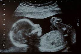
Nt scan
Nuchal Translucency scan in Nagpur: This is actually the prenatal ultrasound test carried out of between weeks 11 or 14 of gestation for the checking chromosomal conditions such as Down syndrome.
Procedure followed at the time of NT scan:
1. Nuchal fold thickness (NT): The sonographer makes an examination of the fluid-filled space behind the neck of the baby.
2. Evaluation of fetal anatomy: The scan also tests the fetal anatomy- head, brain, spine, and limbs.
3. Recognition of any deviation: The sonographer may identify all the visible deviations and abnormalities.
NT Scan (Nuchal Translucency Scan)
Purpose:
– Screen for risk of chromosomal anomalies, such as Down syndrome
– Evaluate fetal anatomy or identify possible abnormalities
– Recommend additional testing and counseling
What to Expect
– 15 to 30 minutes in length
– You may have this during a transabdominal ultrasound, done with the probe placed on your abdomen
– In some cases, you will have a transvaginal ultrasound, which places the probe in your vagina. This is often used in conjunction with the former.
– Sonographer will make a note of the NT’s thickness and check the anatomy of the fetus
– Images or results will be available to you
NT Thickness Fetal Anatomy: Head and brain Spine and limbs Heart and lungs Identification of abnormalities/abnormalities Risk Evaluation Estimated risk for chromosomal abnormalities like Down syndrome Risk levels Low risk Intermediate risk High risk What happens next Care/follow-up Low risk Intermediate or high risk Continue evaluation by further testing e.g. amniocentesis and CVS, or by consulting a specialist e.g. maternal fetal medicine, genetics
What Is a Nt scan?
A Nuchal Translucency (NT) scan is a prenatal ultrasound test performed at 11 to 14 weeks of gestation that evaluates the risk for chromosomal abnormalities, such as Down syndrome, and assesses the anatomy of the fetus. The test specifically measures the thickness of the nuchal fold, that is the fluid-filled space at the back of the fetal neck.
Screens for chromosomal abnormalities, such as Down syndrome, Trisomy 13, and Trisomy 18
It assesses for fetal anatomy and detects anomalies.
It gives recommendations on further testing or consultation based on risk assessment.
A transabdominal scan is typically performed; however, if required, it can also be performed as a transvaginal scan.
The NT scan is the most important tool in:
Assessing early risks and detecting possible chromosomal abnormalities
Guiding further tests and consultations
Providing reassurance and giving a peaceful mind to expectant parents
Purpose of an NT Scan:
- Screen for chromosomal abnormalities: The thickness of the nuchal translucency can indicate a higher risk for certain conditions, including:
- Down syndrome (Trisomy 21).
- Trisomy 18.
- Trisomy 13.
- Detect structural defects: The scan can sometimes detect early signs of heart defects and other structural issues.
- Confirm gestational age: The scan can help confirm how far along the pregnancy is by measuring the baby’s crown-rump length (CRL).
- Check the baby’s heartbeat: The NT scan also assesses the baby’s heart rate and ensures that the baby is developing normally.
How NT Scan Results Are Interpreted:
- Normal range: A nuchal translucency measurement of less than 3.5 mm is generally considered normal, though the exact cutoff may vary slightly depending on the healthcare provider.
- Increased risk: A measurement above the normal range may indicate a higher risk of chromosomal abnormalities, and additional tests, such as a Non-Invasive Prenatal Test (NIPT) or amniocentesis, may be recommended to confirm any concerns.
What is procedures of Nt scan?
Procedural Steps for the NT Scan are as given below:
1. Preparation
– Special preparations are not required
– If it is recommended, go for the test after the full bladder
2. Transabdominal Ultrasound
– The sonographer may use gel or oil at times to enhance the quality of images captured
3. Transvaginal Ultrasound (only if required)
– For better pictures of the fetal anatomy
4. NT Measurement or Nuchal Fold Thickness
– The sonographer measures the thickness of a nuchal fold.
– This measurement is undertaken between 11 to 14 weeks of gestation.
5. Scanning of Fetal Anatomy:
– The sonographer scans the anatomy of a fetus, including:
– Head and Brain
– Spine and Limbs
-Heart and Lungs
6. Detection of Abnormalities:
– A sonographer can possibly see some anomalies or abnormalities.
7. Risk Calculation
– The NT measurement is used for chromosomal abnormality risk calculation
8. Reporting
– A report is issued containing findings and risk evaluation
9. After Scan
– No special post-procedure care is required after scanning
-With you able to resume your normal exercises immediately
At our Neurosys Multispeciality Center, we perform several key procedures including Craniotomy, which is primarily for the excision of brain tumors; V-P Shunt Surgery for treating hydrocephalus; surgeries for epilepsy; and operations targeting brain stem glioma. Beyond these, we offer a range of other neurosurgical services. If you have any questions that are not answere, please contact us through our Contact Us or Book your Appointment.
