
Advanced Assessment Of Fetal Anatomy with Dr.Ritesh Nawkhare
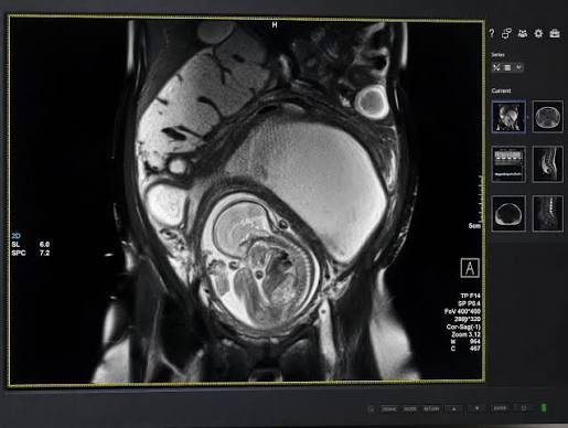
Fetal Magnetic Resonance Imaging (MRI) In Nagpur provides detailed imaging of the developing baby when ultrasound does not provide enough information. Fetal MRI uses magnetic fields and does not use ionising radiation. Doctors may recommend it when they need a clearer view of specific fetal structures or when an ultrasound shows a complex finding. The examination can provide additional information about the fetal brain, spine, chest, abdomen, and other areas, depending on the clinical concern. At Neurosys Multispeciality Centre in Nagpur, fetal imaging concerns can be reviewed under the guidance of Dr. Ritesh Nawkhare. The team considers the gestational age, maternal health, pregnancy history, previous ultrasound findings, and reason for further imaging. Doctors use the MRI findings along with other clinical information rather than assessing the scan in isolation. This approach helps them understand the fetal condition more clearly and decide whether further monitoring, specialist consultation, or additional assessment may be required. The goal is to support informed pregnancy care while keeping maternal and fetal wellbeing at the centre of the evaluation.
What Is Fetal Magnetic Resonance Imaging?
Fetal MRI is an advanced imaging test that uses a magnetic field and radio waves to create detailed images of the developing fetus. It can provide clearer views when ultrasound findings are uncertain, complex, or limited by visibility.
The examination may provide useful information about:
▪ Fetal brain and neurological structures
▪ Spine and surrounding anatomy
▪ Chest and developing lungs
▪ Abdominal organs
▪ Pelvic structures
▪ Selected complex fetal abnormalities
Doctors select the areas to examine based on the reason for the scan. The imaging plan may also vary according to the stage of pregnancy and the clinical findings.
Why Is Fetal MRI Performed?
Ultrasound is the primary imaging method used during pregnancy. However, doctors may not always get clear views of every fetal structure. Fetal position, maternal factors, limited acoustic visibility, or the location of a suspected abnormality can affect the ultrasound assessment.
Doctors may consider Fetal MRI when they need more detailed information about an identified or suspected finding.
Common clinical situations include:
▪ An unusual finding on a detailed ultrasound
▪ Suspected abnormalities of the fetal brain
▪ Possible spinal or neurological conditions
▪ Complex chest or lung findings
▪ Abdominal or pelvic abnormalities
▪ Difficulty obtaining clear ultrasound views
▪ Need for additional anatomical information for further planning
The decision to perform Fetal MRI depends on the clinical findings and the information doctors need to assess the pregnancy more clearly.
Detailed Assessment Of The Fetal Brain
The fetal brain develops continuously throughout pregnancy. When an ultrasound shows a possible neurological concern, MRI can provide additional anatomical detail. It can also help doctors assess structures that are difficult to visualise clearly with ultrasound alone.
Depending on the clinical indication, fetal brain MRI may provide information about:
▪ Cerebral anatomy
▪ Ventricular system
▪ Posterior fossa
▪ Midline structures
▪ Developing brain tissue
▪ Selected suspected developmental abnormalities
Doctors interpret the MRI findings alongside ultrasound observations and other relevant pregnancy information. This combined assessment helps provide a clearer understanding of the fetal brain and any suspected neurological findings.
Fetal Spine And Central Nervous System Imaging
A suspected spinal abnormality may need closer assessment of the spine and surrounding structures. Fetal MRI can provide additional anatomical information when ultrasound findings are complex or incomplete.
The imaging may help clinicians understand:
▪ The location of a suspected finding
▪ The relationship between nearby structures
▪ Associated anatomical features
▪ Whether additional assessment may be useful
Fetal MRI does not provide a diagnosis on its own. Doctors consider the MRI findings along with ultrasound results and other relevant clinical information. This approach supports a more complete assessment of suspected fetal spinal conditions.
Fetal Chest And Lung Assessment For Fetal Magnetic Resonance Imaging (MRI) In Nagpur
Some fetal chest abnormalities can be difficult to assess fully with ultrasound. Fetal MRI can provide additional information about the developing lungs, chest cavity, and nearby structures.
Depending on the clinical indication, the examination may help assess:
▪ Lung development
▪ Chest anatomy
▪ Fluid collections
▪ Space-occupying lesions
▪ Surrounding fetal structures
Doctors use these findings along with ultrasound results and other pregnancy information. The assessment can support ongoing pregnancy monitoring and, when needed, help the medical team prepare for care after birth.
Abdominal And Pelvic Fetal Imaging with Fetal Magnetic Resonance Imaging (MRI) In Nagpur
Fetal MRI can provide additional information when an ultrasound identifies a complex abdominal or pelvic finding. Its wider field of view can help doctors assess the relationship between different anatomical structures.
The assessment may include:
▪ Abdominal organs
▪ Bowel and surrounding structures
▪ Kidneys and urinary system
▪ Liver and adjacent anatomy
▪ Pelvic structures
▪ Fluid-related findings
Doctors select the areas for imaging based on the clinical concern. The imaging plan may vary according to the ultrasound findings and the stage of pregnancy.
When Ultrasound Findings Need More Clarity ?
An ultrasound may identify a finding but may not provide enough detail to fully characterise it. In such cases, fetal MRI can provide additional anatomical information.
It may help doctors assess:
▪ The extent of a suspected abnormality
▪ Its location within the fetal anatomy
▪ Its relationship with nearby structures
▪ Structures that were difficult to visualise on ultrasound
▪ Information that may guide follow-up planning
Advanced imaging aims to answer a specific clinical question. Doctors use the findings along with ultrasound results and other pregnancy information. This supports appropriate monitoring and pregnancy care.
Understanding Fetal MRI Findings with Fetal Magnetic Resonance Imaging (MRI) In Nagpur
Fetal MRI reports may contain specialized medical terms that can be difficult to understand without clinical context. Discussing the findings with an experienced doctor can help parents understand what the scan shows and how it may relate to ongoing pregnancy care. During the consultation, doctors may discuss the findings in relation to:
▪ Previous ultrasound results
▪ Gestational age
▪ The suspected condition
▪ Other pregnancy-related findings
▪ Recommended monitoring
▪ Possible next steps
This discussion helps families understand the available information more clearly. It also allows them to discuss recommended follow-up and pregnancy care with their medical team.
Benefits Of Fetal Magnetic Resonance Imaging (MRI)
Fetal MRI can provide additional anatomical information when ultrasound findings need further clarification. Its potential benefits may include:
▪ Detailed Fetal Imaging – Provides clear views of selected fetal structures.
▪ Advanced Brain Assessment – Provides additional information about fetal brain anatomy when a neurological concern is suspected.
▪ Better Structural Evaluation – Helps assess the spine, chest, lungs, abdomen, and pelvis in selected cases.
▪ Clarifies Complex Findings – Provides additional information when ultrasound results are unclear or difficult to interpret.
▪ No Ionising Radiation – Uses magnetic fields and radio waves instead of X-rays.
▪ Supports Pregnancy Planning – Provides information that can help doctors plan appropriate monitoring and specialist care.
▪ Complements Ultrasound – Adds information to routine prenatal imaging rather than replacing ultrasound.
Recovery After Fetal Magnetic Resonance Imaging (MRI) In Nagpur
After the examination, most patients can return to their usual activities. Fetal MRI does not usually require rehabilitation or special post-procedure care.
After the scan:
▪ Resume Normal Activities – Continue your usual daily routine unless your doctor advises otherwise.
▪ Follow Medical Instructions – Follow any advice provided by your healthcare team based on your pregnancy and clinical needs.
▪ Review the Findings – Discuss the MRI report with your doctor to understand the findings and their relevance to your pregnancy.
▪ Continue Pregnancy Monitoring – Attend scheduled prenatal visits, ultrasound scans, and other recommended assessments.
▪ Attend Follow-Up Appointments – Your doctor may recommend further consultation or imaging when the findings need additional evaluation.
▪ Discuss Further Care – If the scan shows a suspected fetal condition, your doctor can explain the recommended monitoring and possible next steps.
The appropriate follow-up depends on the reason for the MRI and how the findings relate to the overall pregnancy assessment.
Is Fetal MRI Safe During Pregnancy?
Fetal MRI does not use ionising radiation such as X-rays. It uses magnetic fields and radio waves to create detailed images of selected fetal structures. Doctors recommend the examination when they need additional information after reviewing the ultrasound and clinical findings.
Key considerations include:
▪ No Ionising Radiation – MRI uses magnetic fields and radio waves instead of X-rays.
▪ Detailed Fetal Imaging – Provides additional views of selected fetal structures.
▪ Complements Ultrasound – Adds information to ultrasound findings rather than replacing them.
▪ Assesses Complex Concerns – Can help evaluate selected findings that need further anatomical detail.
▪ Individualised Use – Doctors recommend MRI according to the needs of each pregnancy.
▪ Supports Follow-Up – The findings can help doctors plan appropriate monitoring and further assessment.
Your doctor can explain why fetal MRI is recommended, what the examination may assess, and how the findings can support your pregnancy care.
Why Choose Dr. Ritesh Nawkhare For Fetal MRI In Nagpur?
Dr. Ritesh Nawkhare follows a structured and patient-focused approach when reviewing complex pregnancy-related imaging concerns. He considers the available imaging findings along with the overall pregnancy history and clinical context.
His approach may include:
▪ Detailed review of ultrasound and MRI findings
▪ Individualised assessment based on the pregnancy
▪ Careful review of fetal neurological and structural findings
▪ Comparison with previous imaging when available
▪ Clear communication about the reported findings
▪ Guidance on further evaluation and monitoring
▪ Attention to maternal and fetal wellbeing
This approach helps families understand the available imaging information and the recommended follow-up more clearly.
Frequently Asked Questions - Fetal Magnetic Resonance Imaging (MRI) In Nagpur
Book a Consultation for Fetal magnetic resonance (MRI) In Nagpur
If your ultrasound has identified a fetal concern or your doctor has recommended further imaging, consult Dr. Ritesh Nawkhare for a detailed review and personalised guidance.
▪ Review previous ultrasound and pregnancy reports
▪ Discuss the reason for fetal MRI
▪ Understand the areas that may require detailed assessment
▪ Review relevant fetal imaging findings
▪ Plan appropriate follow-up based on the clinical findings