Neuroimaging
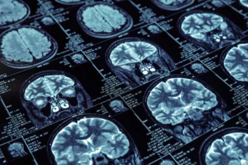
Neuroimaging In Nagpur is the medical area in which the brain is researched. Thus, This is not only used to diagnose conditions and also for evaluation purposes of brain health but also study on how brain functions and how various activities have effects on it.
The NCPRC uses a neuroimaging technique called MRS. Whereas MRI is able to visualize for one the anatomy of the brain, MRS does provide biochemical information. It is sort of like changing the dial on a radio receiver to scan into other chemical nuclei in the brain. We use proton and phosphorus coils in our research in which we explore brain chemistry related to mood disorders in teenagers.
Neuroimaging techniques
Here is the list of some neuroimaging and diagnostic techniques with a better description:
- Positron Emission
- Tomography (PET)
- Magnetic Resonance
- Imaging (MRI)
- Electroencephalography (EEG)
- Magnetoencephalography (MEG)
- Functional MRI (fMRI)
- Computed Tomography (CT)
- Diffusion MRI
- Single-Photon Emission
- Computed Tomography (SPECT)
- Near-Infrared
- Spectroscopy (NIRS)
- Advanced Neuroimaging
- Techniques
- Arterial Spin Labeling (ASL)
- Cranial Ultrasound
- Diagnostic Radiology
- Diffuse Optical Imaging
- Brain Stimulation
- Techniques
- PET Scanning (Repeated for clarity)
Types of Neuroimaging Techniques
- Structural Neuroimaging:
- Magnetic Resonance Imaging (MRI):
- Description: Uses strong magnetic fields and radio waves to create detailed images of the brain and spinal cord.
- Applications: Ideal for detecting tumors, structural abnormalities, and demyelinating diseases (e.g., multiple sclerosis). MRI can also provide insights into brain anatomy and assess conditions like stroke and trauma.
- Advanced Techniques:
- Diffusion Tensor Imaging (DTI): A type of MRI that visualizes white matter tracts and assesses connectivity between different brain regions.
- Magnetic Resonance Angiography (MRA): A specific MRI technique used to visualize blood vessels in the brain.
- Computed Tomography (CT) Scan:
- Description: Utilizes X-rays taken from various angles and processed to create cross-sectional images of the brain.
- Applications: Frequently used in emergency settings to detect acute conditions such as hemorrhages, fractures, and tumors. CT scans are quicker than MRI and are often the first imaging modality used.
- X-ray:
- Description: While not typically used for imaging the brain, X-rays can help visualize the skull and identify fractures or other bony abnormalities.
- Magnetic Resonance Imaging (MRI):
- Functional Neuroimaging:
- Positron Emission Tomography (PET):
- Description: Involves injecting a radioactive tracer into the bloodstream, which emits positrons detectable by the scanner. PET images show areas of metabolic activity in the brain.
- Applications: Useful for assessing brain function, detecting tumors, and evaluating conditions like Alzheimer’s disease and other neurodegenerative disorders.
- Functional Magnetic Resonance Imaging (fMRI):
- Description: A non-invasive technique that measures brain activity by detecting changes in blood flow (hemodynamic response). Active brain regions require more oxygen, which can be visualized through fMRI.
- Applications: Widely used in research and clinical settings to study brain function, assess cognitive processes, and plan neurosurgical interventions. fMRI is valuable in mapping critical brain functions (e.g., language, motor function) before surgery.
- Single Photon Emission Computed Tomography (SPECT):
- Description: Similar to PET but uses different radioactive tracers. SPECT provides images of blood flow and metabolic activity in the brain.
- Applications: Often used in the evaluation of dementia, seizure disorders, and certain psychiatric conditions.
- Positron Emission Tomography (PET):
- Electrophysiological Techniques:
- Electroencephalography (EEG):
- Description: Measures electrical activity in the brain through electrodes placed on the scalp. It provides real-time information about brain function.
- Applications: Primarily used to diagnose epilepsy, sleep disorders, and other neurological conditions. EEG is also valuable in monitoring brain activity during surgery.
- Magnetoencephalography (MEG):
- Description: Measures the magnetic fields generated by neuronal activity. It provides excellent temporal resolution, allowing for real-time monitoring of brain function.
- Applications: Useful for localizing brain functions prior to surgery and understanding brain activity patterns in various cognitive tasks.
- Electroencephalography (EEG):
Applications of Neuroimaging
Diagnosis: Neuroimaging is crucial for diagnosing a wide range of neurological conditions, including strokes, tumors, traumatic brain injuries, neurodegenerative diseases (e.g., Alzheimer’s), and psychiatric disorders.
Treatment Planning: Imaging helps neurosurgeons plan procedures by mapping critical brain areas and identifying the precise location of lesions.
Research: Neuroimaging is essential for understanding brain structure-function relationships, cognitive processes, and the effects of interventions (e.g., medication, therapy).
Monitoring: Follow-up imaging can assess disease progression, treatment response, and the effectiveness of interventions.
Limitations and Considerations
Cost and Accessibility: Neuroimaging techniques, particularly MRI and PET, can be expensive and may not be available in all healthcare settings.
Radiation Exposure: CT and PET involve exposure to radiation, necessitating careful consideration, particularly in vulnerable populations like children.
Interpretation: Neuroimaging requires specialized training for accurate interpretation, as normal variants and incidental findings can sometimes be misinterpreted as pathological.
Clinical Context: Imaging findings should always be interpreted in conjunction with clinical history and symptoms to guide appropriate management.
Clinical Applications of Neuroimaging:
- Diagnosis:
Neuroimaging plays the critical role in a diagnosing neurological disorders, including:
Stroke: CT and MRI can identify ischemic or hemorrhagic strokes and guide treatment decisions.
Tumors: Imaging helps locate and characterize brain tumors, allowing for appropriate treatment planning.
Neurodegenerative Diseases: MRI and PET can assist in diagnosing conditions like Alzheimer’s disease, Parkinson’s disease, and multiple sclerosis. - Treatment Planning:
Neuroimaging aids in surgical planning for brain surgeries, including tumor resections and deep brain stimulation. - Monitoring Disease Progression:
Imaging can track changes in brain structure or function over time, providing valuable information on disease progression and treatment efficacy. - Research:
Neuroimaging is essential in neuroscience research, helping to explore brain connectivity, neural mechanisms underlying behaviors, and the effects of various interventions (e.g., medications, therapies).
What is brain imaging used for?
Brain imaging is highly crucial in the healthcare sector because it gives doctors ample opportunities to make proper diagnoses. Long ago, Some of the important applications of brain imaging techniques are as follows:
- Determine the effects of a stroke.
- Detection of cysts and tumors
- Identify areas that are swollen and heavy with blood.
Hence, the selection of imaging technique should depend on specific diagnostic needs. For instance, if the symptoms had suggested MS, the physician would refer the patient for MRI either to confirm or dismiss MS lesions. However, in a situation like investigating a case of bone fracture, a CT scan would be better suited since it would give clearer images of the internal bones’ structure.
